
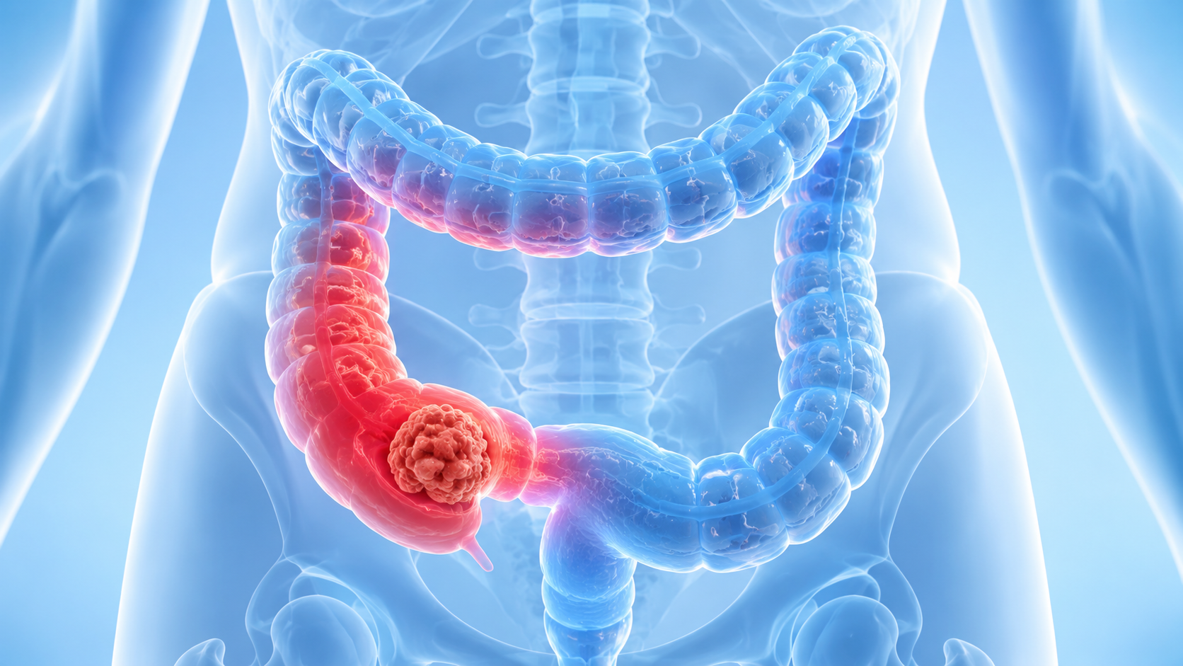
Chinese Expert Whole-Process Management Guidelines for Early-Stage Colorectal Cancer
Colorectal Cancer: The Earlier the Detection, the More Treatment Options
Colorectal cancer is one of the most common malignancies worldwide, ranking third in global incidence and second in mortality.
Many patients, upon hearing a diagnosis of colorectal cancer, immediately think of major surgery, chemotherapy, stoma, and lengthy treatment. However, for early-stage colorectal cancer, the situation is not entirely the same. If the tumor is detected sufficiently early, treatment approaches may differ significantly, and some patients may even be treated with endoscopic resection or local excision.
According to the Chinese Guidelines for Whole-Process Management of Early-Stage Colorectal Cancer, early-stage colorectal cancer refers to cancer confined to the mucosal and submucosal layers of the colorectum, meaning the cancer cells have not yet invaded the deeper muscularis propria. It primarily corresponds to Tis and T1 stage colorectal cancer.
The shallower the depth of tumor invasion, the lower the risk of lymph node metastasis generally is, and the greater the opportunity for patients to receive minimally invasive treatment, local therapy, or function-preserving treatment.
For international patients, a second opinion after early diagnosis is very important. Many patients are found to have polyps, adenomas, high-grade intraepithelial neoplasia, or early cancer during colonoscopy in their home country but are unclear about the next steps. Some patients may be advised to undergo surgery directly, while others may be suitable for more refined endoscopic evaluation, pathological review, and imaging assessment first.
Major digestive endoscopy centers and colorectal surgery centers in China have established a complete diagnosis and treatment system for early-stage colorectal cancer, including diagnosis, endoscopic treatment, laparoscopic/robotic surgery, and multidisciplinary consultation.
Evaluation of Early-Stage Colorectal Cancer
The evaluation of early-stage colorectal cancer typically includes: colonoscopy, pathological diagnosis, MMR/MSI testing, contrast-enhanced CT or MRI, etc.
Colonoscopy is the core tool for colorectal cancer screening and diagnosis; contrast-enhanced abdominal CT is recommended for early colon cancer evaluation, and contrast-enhanced pelvic MRI is recommended for early rectal cancer. For rectal cancer patients, MRI is particularly important because it helps determine tumor location, depth of invasion, relationship with surrounding structures, and suspicious lymph nodes.
If a patient has already completed initial examinations overseas and seeks medical advice in China, it is generally recommended to prepare the following materials: colonoscopy report, colonoscopy images or video, pathology report, pathology slides or paraffin blocks, CT/MRI imaging data, tumor marker results, and previous treatment records. Doctors need to re-evaluate whether the lesion is truly early-stage, whether high-risk factors exist, and whether there is an opportunity for more minimally invasive treatment.
Treatment Goals for Early-Stage Colorectal Cancer
In addition to tumor eradication, the goals are to minimize trauma, preserve bowel function, reduce recurrence risk, and improve long-term quality of life.
For some patients, early detection means avoiding extensive surgery; for others, even if surgery is necessary, a more appropriate surgical extent and timing can be selected through standardized evaluation.
Therefore, if a patient is diagnosed with Tis or T1 stage colorectal cancer, high-grade intraepithelial neoplasia, or pathology after endoscopic resection indicates carcinoma, it is recommended to seek comprehensive evaluation from an experienced colorectal cancer team as soon as possible. The earlier the risks are clarified, the greater the opportunity to choose a treatment plan that is more precise, less traumatic, and more aligned with quality of life needs.
EMR, ESD, and Local Excision
The most common concern for many early-stage colorectal cancer patients is: Will it be major surgery? Can the anus and normal defecation function be preserved? The answer is yes.
The reason early-stage colorectal cancer has the opportunity for minimally invasive treatment is that the tumor is still confined to the mucosal or submucosal layer. The basic indication for endoscopic resection of early-stage colorectal cancer is: low risk of lymph node metastasis, and based on tumor size and location, it is expected that complete endoscopic resection can be achieved. In other words, not all "early cancers" can be treated endoscopically; the key is whether there is a risk of lymph node metastasis and local residual disease.
Currently, common endoscopic and local treatment methods include EMR, ESD, transanal local excision, TEM, and TAMIS.
EMR is Endoscopic Mucosal Resection
It is suitable for some flat or sessile lesions that can be completely resected in one piece. In principle, the maximum diameter of lesions that can be resected en bloc by EMR is usually less than 20mm. For appropriate lesions, EMR causes less trauma and allows faster recovery, making it an important method in the treatment of early lesions.
ESD is Endoscopic Submucosal Dissection
It is suitable for more complex early lesions. Compared with EMR, the main advantages of ESD are a higher en bloc resection rate and a lower recurrence rate. It is particularly suitable for lesions with a maximum diameter exceeding 20mm, those difficult to resect completely in one piece by EMR, those with a negative lifting sign, and those with residual or recurrent lesions after EMR where repeat EMR is difficult.
For lesions highly suspicious of superficial submucosal invasion based on morphology, ESD also has significant value. However, ESD requires longer procedure time, carries a higher risk of complications, and demands greater technical skill from the endoscopist.
Local Excision
For mid-to-low early-stage rectal cancer, transanal local excision can also be considered. Common methods include traditional transanal local excision, TEM, and TAMIS.
TEM is transanal endoscopic microsurgery, and TAMIS is transanal minimally invasive surgery. Their value lies in the ability to resect some early rectal lesions via the anal approach, avoiding or reducing the trauma associated with major abdominal surgery. For early-stage rectal cancer with a low location, lesions meeting low-risk criteria, and patients who strongly desire to preserve anal function, local excision may be a treatment option worth evaluating.
Minimally Invasive Does Not Mean the Smaller the Better
Minimally invasive treatment is not about being as small as possible; it is about minimizing trauma while ensuring safety. If pathology indicates higher risk, such as poor differentiation, lymphovascular invasion, perineural invasion, deep submucosal invasion, or positive margins, endoscopic resection or local excision alone may be insufficient. In such cases, ignoring the risk of lymph node metastasis in an effort to "cut less" may actually increase the risk of recurrence and metastasis.
This is why major colorectal cancer centers in China typically emphasize multidisciplinary evaluation. The treatment of early-stage colorectal cancer requires the digestive endoscopist to determine whether complete resection is feasible, the pathologist to assess the risk level, the radiologist to evaluate lymph nodes and local invasion, and the colorectal surgeon to determine whether additional surgery is needed. For patients with low rectal cancer, it is also necessary to focus on sphincter preservation, defecation function, and quality of life.
Guide for International Patients
For international patients who have been diagnosed with early-stage colorectal cancer in their home country but are unsure whether major surgery is necessary, they may consider seeking a second opinion from Chinese experts with complete medical records. The following situations especially warrant re-evaluation: the lesion is relatively large but it is unclear whether ESD resection is possible; the doctor recommends radical surgery but the patient wishes to know if local treatment is an option; early-stage rectal cancer patients worried about permanent stoma; complex pathology results after endoscopic resection requiring judgment on whether additional surgery is needed.
The ideal state of early-stage colorectal cancer treatment is to minimize trauma and preserve organ function and quality of life while ensuring oncological safety. EMR, ESD, TEM, and TAMIS are not simple interchangeable options but precise treatment tools for different lesions, risks, and locations.
Additional Surgery May Be Necessary
Many patients, after having polyps or early cancer removed endoscopically, think the treatment is complete. However, upon receiving the pathology report, the doctor recommends additional surgery. Patients are often confused: hasn't the tumor already been removed? Why is more surgery needed?
The reason is that endoscopic resection addresses the local lesion but cannot clear lymph nodes. If pathology indicates a risk of lymph node metastasis, local residual disease, or incomplete resection, the patient may need additional radical surgery.
Main Indications for Additional Surgery
Positive horizontal margin, meaning cancer tissue is less than 1mm from the resection margin, or tumor cells are present at the margin; positive vertical margin; depth of submucosal invasion ≥1000μm; presence of lymphovascular invasion or perineural invasion; poor tumor differentiation; tumor budding G2 or G3; incomplete resection, fragmented specimen, unevaluable margins, or unclear pathological findings, etc.
A positive margin indicates that tumor cells may be close to or have reached the resection edge, with a higher risk of local residual disease.
Submucosal invasion depth ≥1000μm indicates that cancer cells have invaded deeper layers, increasing the risk of lymph node metastasis.
Lymphovascular invasion means cancer cells have entered blood vessels or lymphatic vessels, and perineural invasion means cancer cells have invaded tissues around nerves; these are signals of a higher risk of tumor spread.
Pathological types such as poorly differentiated, undifferentiated, mucinous adenocarcinoma, and signet ring cell carcinoma usually have more unstable biological behavior.
Tumor budding G2 or G3 indicates more aggressive tumor behavior and is also associated with a higher risk of lymph node metastasis.
Additional Surgery Does Not Mean Endoscopic Treatment Was Meaningless
Because the pathology results after endoscopic treatment provide more precise risk information, many early-stage colorectal cancer patients clarify whether additional radical surgery is needed precisely through initial endoscopic resection followed by pathological evaluation.
For colon cancer, if the indication for additional surgery is clear, it is recommended to complete the additional surgery within 4–8 weeks as soon as possible; for rectal cancer, it should be completed as soon as possible after ensuring surgical safety, taking into account the patient's condition and local wound status.
Radical surgery is generally recommended for additional surgery. For patients with low rectal cancer, comprehensive treatment options such as radiotherapy may also be chosen based on clinical circumstances and patient preference, but risks must be fully assessed.
Guide for International Patients
For international patients, the post-endoscopic treatment phase is very suitable for a second opinion. Because whether additional surgery is needed requires careful review of the pathology report and resected specimen. Some patients do need additional radical surgery; others may have low-risk complete resection and can be closely followed up.
If the patient has already undergone endoscopic resection, it is recommended to prepare the following materials: original colonoscopy report, endoscopic images or video, complete pathology report, pathology slides or paraffin blocks, CT/MRI images, and tumor markers such as CEA.
If the pathology report contains terms such as "positive margin," "submucosal invasion," "lymphovascular invasion," "perineural invasion," "poor differentiation," "tumor budding," or "fragmented specimen," it is especially advisable to seek evaluation from an experienced colorectal cancer team.
Major hospitals in China have a complete multidisciplinary collaboration model in digestive endoscopy, colorectal surgery, pathological review, and imaging evaluation. For international patients who have received initial treatment but whose treatment path is unclear, the Chinese expert team can help determine: whether additional surgery is needed, the extent of surgery, whether it can be performed minimally invasively, whether there is a chance for sphincter preservation, and how to follow up subsequently.
Chinese Doctors Evaluate Sphincter Preservation, Function Preservation, and Radical Efficacy
Although both rectal cancer and colon cancer fall under colorectal cancer, the concerns patients have are often different. For rectal cancer, especially mid-to-low rectal cancer, the most common concerns are: Can the anus be preserved? Will there be a permanent stoma? Will postoperative defecation function be affected? Can quality of life be restored?
If early-stage rectal cancer is detected early, some patients do have the opportunity to receive local excision, transanal surgery, or other function-preserving treatments. However, such treatments must be based on strict selection criteria. Not all early-stage rectal cancers are suitable for local excision and permanent stoma avoidance; the key lies in precise evaluation.
Early-stage rectal cancer is highly suitable for multidisciplinary consultation, especially the MDT model involving colorectal surgery, gastrointestinal surgery, pathology, digestive endoscopy, medical imaging, medical oncology, and radiation oncology. In particular, patients with early-stage colorectal cancer with high-risk factors, early low rectal cancer, or unclear T staging are more suitable for treatment decisions through MDT discussion.
Guide for International Patients
For international patients, the value of coming to China for evaluation of early-stage rectal cancer is mainly reflected in three aspects.
Re-evaluating Whether Local Treatment Is Truly Suitable
Some patients are advised to undergo radical surgery overseas, but after more detailed endoscopic, MRI, and pathological evaluation, there may be an opportunity for local excision or more conservative treatment.
Assessing the Possibility of Sphincter Preservation
Treatment of low rectal cancer should not only pursue resection but also consider anal function, defecation function, and postoperative quality of life. For some patients, TEM, TAMIS, transanal local excision, laparoscopic, or robotic surgery may all be part of the treatment plan.
Avoiding Sacrificing Radical Efficacy for Sphincter Preservation
Some patients strongly wish to avoid a stoma, but if there is a risk of lymph node metastasis or pathological high-risk factors, local treatment alone may be unsafe. In such cases, doctors need to balance oncological radicality, sphincter preservation opportunity, recurrence risk, and patient quality of life.
If a patient has been diagnosed with early-stage rectal cancer, it is recommended to prepare the colonoscopy report, colonoscopy images, pathology report, pelvic MRI, chest and abdominal CT, CEA, and other materials for comprehensive evaluation by the colorectal cancer team. For patients who have already undergone local excision or ESD, complete pathology slides should also be provided to determine whether additional treatment is needed. The Chinese colorectal cancer multidisciplinary team will provide you with refined evaluation and treatment options.